In this article, the muscles of the leg are described, following the line of previous publications (the bones of the leg, the different types of joints and the joints of the leg).
What are the muscles of the leg?
The muscles of the leg are distributed in different regions, planes, and compartments and have different functions (1, 2, 3). This means that, although the main function of a muscle is specific (e.g., the muscle group of the quadriceps femoris, extending the knee joint), they can have other functions (e.g., the long flexor muscle of the big toe flexes the big toe, but also assists in plantar flexion of the foot and plays an important role in the toe-off phase during walking) (1).
[article ids=”80461″] Thus, in this article, the different muscles of the leg are described, distributed by the various regions, planes, and compartments of the lower limb, in an order from proximal to distal and from deep to superficial, also specifying their points of origin and insertion, as well as the actions they perform and help perform.
*Note: in most of the images in this article, it is not specified whether it is an anterior or posterior view because a large part are oblique views, seeking better visualization of the insertions of the leg muscles.
Gluteal region
The gluteal region is the posterior transition zone between the trunk and the lower limb and is formed by the hip region and the buttocks (2).
It is located in the posterior part of the body, forming the postero-lateral-inferior part of the pelvis, from the iliac crests to the posterior edge of the greater trochanter (1, 2).
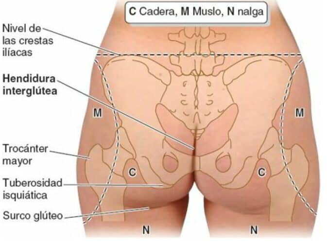
This region is connected to the pelvic cavity through the greater sciatic foramen and to the perineum through the lesser sciatic foramen (1).
Deep plane
In this plane, there are small leg muscles that are mainly external rotators of the hip joint (1).
Piriformis/pyramidal
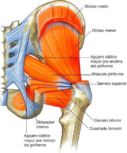
The piriformis/pyramidal muscle is a leg muscle that belongs to both the gluteal region and the pelvic wall (1, 2). It has a rectilinear path (3) with an infero-lateral direction (1).
From its point of origin, it reaches its insertion through the greater sciatic foramen (1, 2). The piriformis/pyramidal muscle is also an anatomical reference because it divides the greater sciatic foramen into 2, one superior and one inferior to the muscle itself, through which vessels and nerves pass (1, 2).
-
-
-
- Origin (1, 2)
-
-
-
- On the surface between the anterior sacral foramina.
-
-
-
-
-
- Sacrotuberous ligament.
-
-
-
- Origin (1, 2)
-
-
-
-
-
- Insertion (1, 2)
Internal area of the superior border of the greater trochanter.
- Insertion (1, 2)
-
-
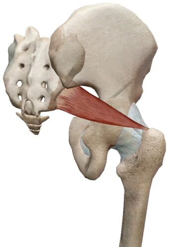
Actions (1, 2):
-
- External rotation of the hip joint in extension.
-
- Abduction of the hip joint in flexion.
Internal obturator
The tendon of the internal obturator rotates 90º (1, 2, 3) around the ischium between 2 bony references (ischial spine and ischial tuberosity) (1), over a synovial bursa (3) and enters the gluteal region through the lesser sciatic foramen, following a postero-inferior path to its insertion (1, 2).
Together with the superior and inferior gemellus leg muscles, it forms the triceps coxae muscle, whose common tendon inserts into the greater trochanter of the femur (2).
[caption id="attachment_80284" align="aligncenter" width="387"]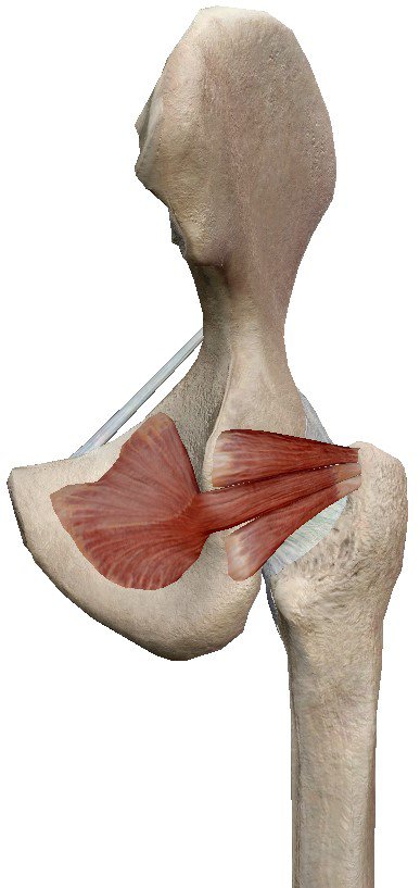
- Origin (1, 2)
- Anterolateral wall of the lesser pelvis.
- Internal surface of the obturator membrane
- Internal surface of the bone of the obturator foramen.
- Insertion (1, 2)
Internal part of the superior border of the greater trochanter, inferior to the insertion of the piriformis/pyramidal muscle through a common tendon with the superior and inferior gemellus leg muscles.
[caption id="attachment_80285" align="aligncenter" width="373"]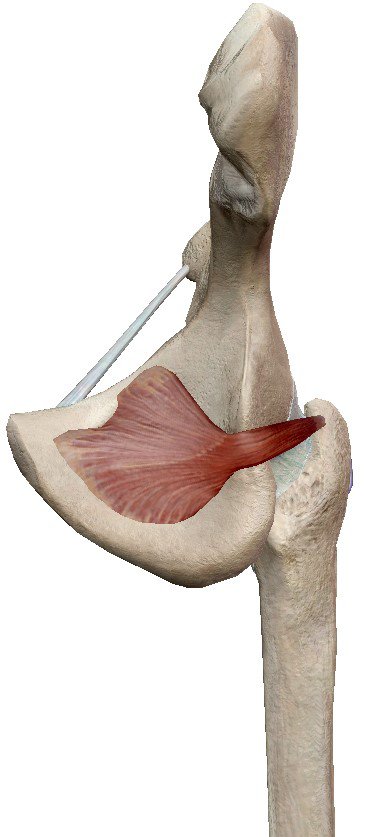
Actions (1, 2):
- External rotation of the hip in extension.
- Abduction of the hip in flexion.
- Stabilization of the femoral head in the acetabulum.
Superior and inferior gemellus
These are 2 triangular-shaped leg muscles that are related to the tendon of the internal obturator muscle, inserting one on its superior surface (superior gemellus) and the other on its inferior surface (inferior gemellus) (1, 2, 3).
- Origin
- External surface of the ischial spine (superior gemellus) (1, 2).
- Superior gluteal surface and pelvic surface of the ischial tuberosity (inferior gemellus) (1, 2).
- Insertion
Internal part of the greater trochanter with the tendon of the internal obturator muscle. They join this tendon along its superior surface (superior gemellus) and its inferior surface (inferior gemellus) (1, 2).
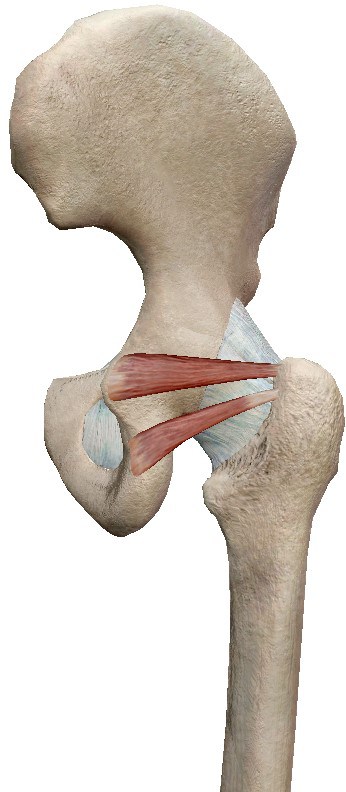
Actions (1, 2):
- External rotation of the hip in extension.
- Abduction of the hip in flexion.
- Stabilization of the femoral head in the acetabulum.
Quadratus femoris
It is a flat, rectangular-shaped leg muscle located immediately inferior to the inferior gemellus, thus being the most inferior of all in this group (1, 2).
- Origin (2)
External border of the ischial tuberosity. - Insertion (1, 2)
Quadrate tubercle of the intertrochanteric crest and area inferior to it.
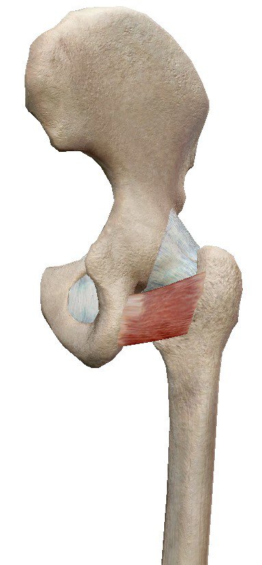
Actions (1, 2):
- External rotation of the hip in extension (powerful).
- Stabilization of the femoral head in the acetabulum.
Superficial plane
The leg muscles located in the superficial plane of the hip are larger than the muscles of the deep plane, mainly abductors and extensors of the hip (1).
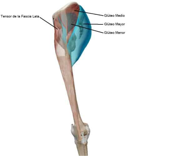
Gluteal muscles
There are 3 gluteal muscles, these leg muscles are superimposed on each other from deep to superficial and from anterior to posterior (1, 3).
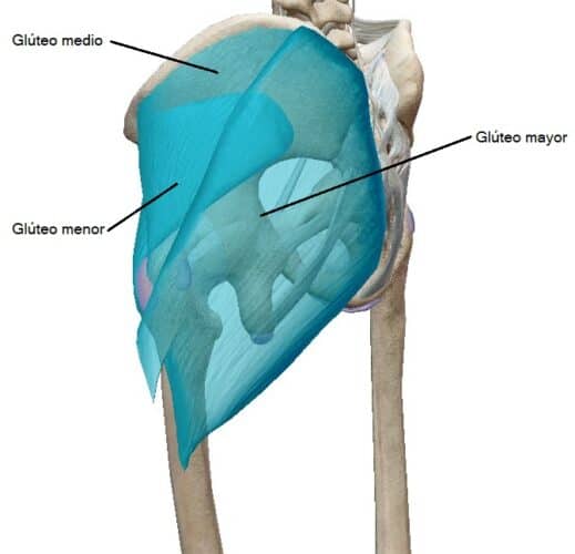
Gluteus minimus and gluteus medius
Both have a fan shape and are located deep to the gluteus maximus muscle, with the gluteus minimus being deeper than the gluteus medius (1, 2).
- Origin (1, 2, 3)
- Gluteus minimus: gluteal surface, between the inferior and anterior gluteal lines.
- Gluteus medius: between the anterior and posterior gluteal lines.
- Insertion (1, 2)
- Gluteus minimus: facet on the antero-external surface of the greater trochanter.
- Gluteus medius: facet on the external surface of the greater trochanter.
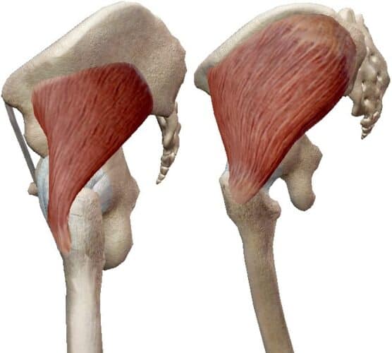
Actions (1, 2, 4, 5):
- Abduction of the hip joint.
- Internal rotation of the hip joint (anterior fibers of the gluteus medius, superior fibers of the gluteus minimus).
- External rotation of the hip joint (posterior fibers of the gluteus medius, inferior fibers of the gluteus minimus).
- Maintain the pelvis steady on the supporting leg.
- Prevent the contralateral pelvis from dropping during walking.
Gluteus maximus
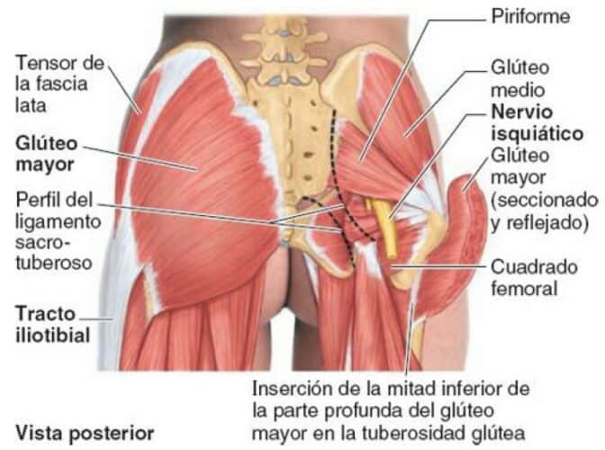
It is the largest gluteal muscle and the most superficial of the 3 gluteal muscles (covering part of the gluteus minimus and medius) (1, 2, 6) and is the muscle with the thickest fibers in the body (2). It has a quadrangular shape, with a very wide area of origin (1, 2).
It is the most powerful extensor of the hip joint, but it mainly acts under power demand or against high resistance (2).
- Origin (1, 2, 3)
- Fascia that envelops the gluteus medius.
- Behind the posterior gluteal line of the gluteal surface.
- Fascia of the erector spinae muscle.
- Posterior surface of the lower part of the sacrum.
- Posterior part of the coccyx.
- External surface of the sacrotuberous ligament.
- Insertion (1, 2, 3)
- On the external surface of the iliotibial band on its posterior side, which inserts into the external tibial condyle.
- On the gluteal tuberosity of the proximal end of the femur.
- Linea aspera.
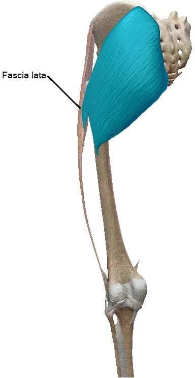
Actions (1, 2):
- Extension of the hip joint from flexion (standing up from a sitting position, climbing stairs…).
- External rotation of the hip joint.
- Lateral stabilization of the hip and knee joints.
Tensor fasciae latae (TFL)
The TFL is, among the leg muscles, the most anteriorly located in the superficial plane of the gluteal region (1, 2, 6). It is located covering the gluteus minimus and the anterior part of the gluteus medius (1, 6).
- Origin (1, 2)
On the lateral part of the iliac crest, from the anterior superior iliac spine to the crest tubercle. - Insertion (1, 2)
On the anterior side of the iliotibial band, which inserts into the proximal end of the tibia. This insertion point is common with the superficial and anterior part of the gluteus maximus muscle.

Actions (1, 2):
- Stabilize the knee in extension.
- Stabilize the hip by fixing the femoral head in it.
- Maintain the pelvis level when the same limb bears the weight.
- The role of the tensor fasciae latae muscle as an abductor and/or internal rotator of the hip joint, as well as in the flexion and external rotation of the knee joint, given its very anterior location in the gluteal region, are probably synergistic muscle actions.
Thigh
The leg muscles located in the thigh are organized into 3 compartments (1, 2):
- Anterior compartment
- Distal ends of the iliacus and psoas major leg muscles.
- Quadriceps
- Sartorius.
- Internal compartment
- External obturator.
- Pectineus.
- Adductor brevis.
- Adductor longus.
- Adductor magnus.
- Gracilis.
- Posterior compartment:
- Hamstrings.
Some authors include the pectineus muscle in the anterior compartment and others in the internal compartment (1, 2), although they detail that due to its dual innervation and dual functionality, it is often considered a transitional muscle between the anterior and internal compartments (2).
In this article, since it is located in the supero-internal part of the thigh, it is included in the internal compartment.
Anterior compartment of the thigh
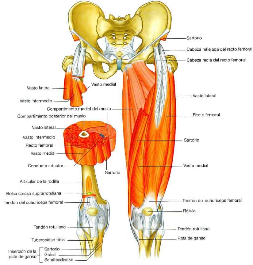
In the anterior compartment of the thigh, there are some of the most voluminous leg muscles (2) and they act on the hip and knee (1).
Iliopsoas/psoas iliacus
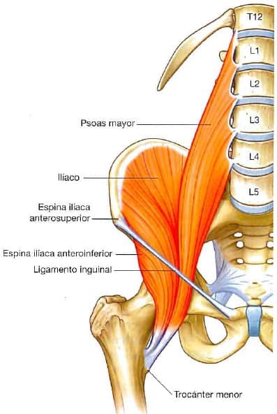
The psoas major and iliacus muscles are 2 leg muscles whose origin occurs at different points on the posterior abdominal wall, but they insert through a common tendon on the trochanter of the femur. Since their distal insertion is on the femur, their action on the hip is considered one of the leg muscles (1, 2).
- Origin (1, 2)
- Lumbar transverse processes, intervertebral discs, and tendinous arches between these points (Psoas major).
- Iliac fossa (Iliacus).
- Insertion (1, 2, 3)
Lesser trochanter of the femur.
Actions (1, 2):
- Hip flexion (powerful).
- Initiates anterior swing during walking.
- Raises the limb in climbing.
- External rotation of the hip joint (assists).
- Flexion of the trunk over the hip joint (fixed lower limb).
- Reduction of lumbar lordosis (fixed lower limb).
- Maintenance of lumbar lordosis (standing).
- Resistance to hyperextension of the hip joint (standing).
Quadriceps
It is formed by 4 heads/portions/vasti, which have different points of origin but unite in a single common tendon (1, 2, 3, 7).
The different portions of the quadriceps muscle are (1, 2, 3, 7):
Vastus intermedius
It is located deep to the rectus femoris, between the vastus medialis and lateralis.
Rectus femoris
Biarticular, very effective in combined movements of hip flexion and knee extension.
Vastus medialis
Occupies the inner part of the thigh
Vastus lateralis
It is located on the outer part of the thigh and occupies the largest volume of the quadriceps muscle.
- Origin (1, 2)
- Vastus intermedius – Upper two-thirds of the anterior and lateral surfaces of the femur.
- Rectus femoris – Anterior inferior iliac spine and ilium, superior to the acetabulum.
- Vastus medialis – Medial part of the intertrochanteric line, pectineal line, medial lip of the linea aspera, and medial supracondylar line.
- Vastus lateralis – Lateral part of the intertrochanteric line, border of the greater trochanter, lateral border of the gluteal tuberosity, and lateral lip of the linea aspera.
- Insertion (1, 2, 3, 7,)
Through the quadriceps tendon (quadriceps tendon) at the base of the patella, as well as on the patella itself and through the patellar ligament extends to the tibial tuberosity. Each portion inserts into a part of the quadriceps tendon and the patella: - Vastus intermedius on the deep surface of the quadriceps tendon and on the lateral border of the patella.
- Rectus femoris in addition to the quadriceps tendon, on the base of the patella.
- Vastus medialis on the medial surface of the quadriceps tendon and on the medial border of the patella.
- Vastus lateralis in addition to the quadriceps tendon, on the lateral border of the patella.
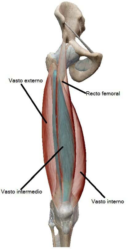
Thus, the rectus femoris is the only portion of the quadriceps muscle that is biarticular, acting on the hip and knee (1, 2, 3, 6). It is the major extensor of the knee joint and can be up to 3 times more powerful than the hamstring leg muscles (antagonistic muscle group) (2).
Actions (1, 2, 7):
- Extension of the knee joint.
- Stabilizes the hip joint (rectus femoris).
- Flexion of the hip joint along with the iliopsoas muscle (rectus femoris).
- Absorption of impact and support of body weight during walking.
Articularis genus muscle
The articularis genus muscle is a small leg muscle derived from the vastus intermedius of the quadriceps muscle (2) and moves the suprapatellar bursa during knee extension (1, 2).
- Origin (1, 6)
On the lower part of the anterior surface of the femur, inferior to the origin of the vastus intermedius. - Insertion (1, 2, 6)
Suprapatellar bursa.
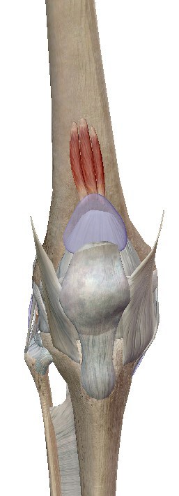
Actions (1, 2):
- Move the suprapatellar bursa of the knee during extension by pulling it.
Sartorius
The sartorius muscle is the longest muscle in the body and therefore also the longest of the leg muscles (2).
From its point of origin, it descends in an inferomedial direction to its point of insertion, crossing the quadriceps muscle in an anterior plane (1, 2, 3, 8).
- Origin (1, 2, 3, 8)
Anterior superior iliac spine (ASIS). - Insertion (1, 2)
Medial surface of the proximal tibial shaft, anterior to the insertions of the gracilis and semitendinosus leg muscles and inferolateral to the tibial tuberosity.

Since the insertion of the sartorius muscle along with the insertions of the gracilis and semitendinosus muscles follows a “three-point” pattern (1), some authors determine that the sartorius muscle inserts into the pes anserinus (1, 2, 3).
Actions (1, 2, 8):
- Flexion of the hip joint (assists).
- Flexion of the knee joint (assists).
- Abduction of the hip joint.
- External rotation of the hip joint if the foot is resting on the opposite knee.
- Allows sitting with crossed legs (bilateral action).
- The role of the sartorius muscle is mainly synergistic.
The sartorius muscle is called “the tailor’s muscle” or simply abbreviated as sartorius, named after the posture tailors used to adopt, crossing their legs when working (2, 8).
Internal compartment of the thigh
In the internal compartment, there are 6 leg muscles, all but 1 primarily adduct the hip (1, 2):
- External obturator.
- Pectineus.
- Adductor brevis.
- Adductor longus.
- Adductor magnus.
- Gracilis.
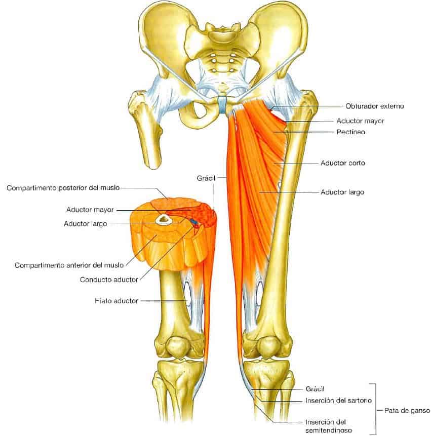
External obturator
The external obturator muscle is a flat, fan-shaped leg muscle that connects the pelvis with the femur, therefore, it acts directly on the hip joint (1, 2).
It is located deep in the proximal and internal part of the thigh, and its tendon passes under the femoral neck to its point of insertion (2, 1).
- Origin (1, 2)
External surface of the obturator membrane and adjacent bone. - Insertion (1, 2)
Depression of the external wall of the trochanteric fossa.
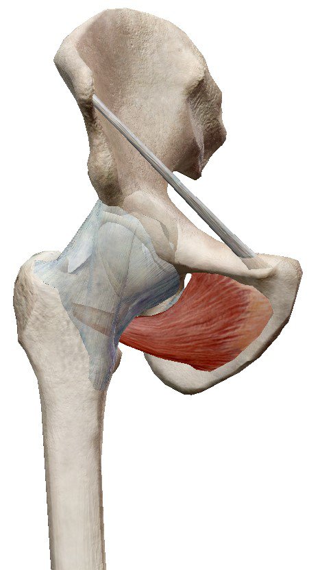
Actions (1, 2):
- External rotation of the hip joint (more effective with the hip joint in flexion).
- Stabilization of the femoral head in the acetabulum.
Pectineus
The pectineus muscle is a flat, quadrangular leg muscle that extends inferolaterally from the pelvis to the proximal end of the femur (1, 2), passing inferiorly to the inguinal ligament (1).
- Origin (1, 2)
Pectineal line of the pelvis. - Insertion (1, 2)
Oblique line from the base of the lesser trochanter to the posterior linea aspera of the proximal end of the femur.
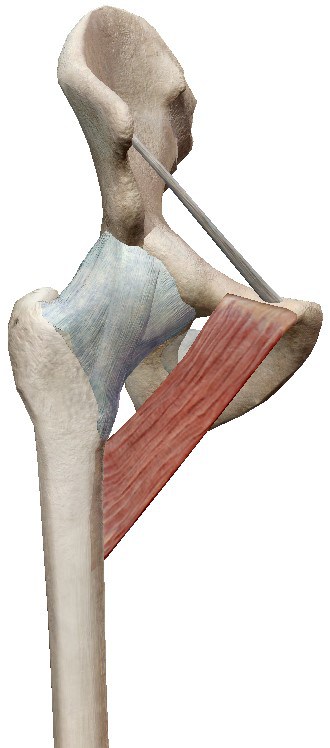
Actions (1, 2):
- Adduction of the hip joint.
- Flexion of the hip joint.
- Internal rotation of the hip joint (assists).
Adductor brevis
The adductor brevis muscle is a triangular-shaped leg muscle (1) located posterior to the pectineus and adductor longus muscles (1, 2).
- Origin (1, 2)
On the body of the pubis and superior to the origin of the gracilis muscle on the inferior pubic ramus. - Insertion (1, 2)
On the pectineal line and the proximal part of the linea aspera of the femur, lateral to the insertions of the muscles.
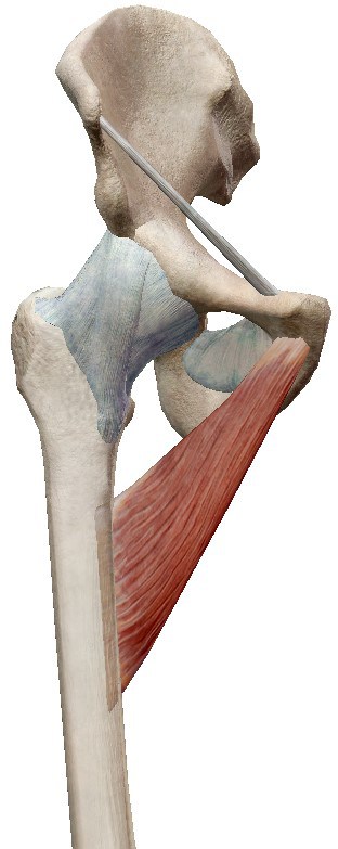
Actions (1, 2):
- Adduction of the hip joint.
- Slight flexion of the hip joint.
Adductor longus
The adductor longus muscle is a large, flat, fan-shaped leg muscle (1, 2). It is the most anteriorly positioned muscle of the leg muscles that adduct the hip joint (2).
- Origin (1, 2)
On a rough triangular surface of the external part of the pubis, inferior to the pubic crest, external to the pubic symphysis. - Insertion
On the middle third of the linea aspera.

From its point of origin, it expands adopting that fan shape and is oriented postero-laterally to its insertion (1, 2).
Actions (1, 2):
- Adduction of the hip joint.
- Internal rotation of the hip joint.
Adductor magnus
The adductor magnus muscle is a triangular, fan-shaped leg muscle and is the largest and deepest of the internal compartment of the thigh (1, 2).
It consists of 2 portions with different insertions, innervation, and (main) actions, the adductor portion (more external) and the ischiotibial part (more internal), between which is the tendinous hiatus or adductor hiatus (1, 2), through which the femoral artery and several veins pass between the adductor canal to the popliteal fossa (1).
- Origin (1, 2)
- Adductor part
On a line from the inferior pubic ramus and on the ischial ramus. - Ischiotibial part
On the ischial tuberosity.
- Adductor part
- Insertion (1, 2)
- Adductor part
On the gluteal tuberosity, along the linea aspera, on the medial supracondylar line. - Ischiotibial part
On the adductor tubercle of the medial femoral condyle and on the medial supracondylar line.
- Adductor part
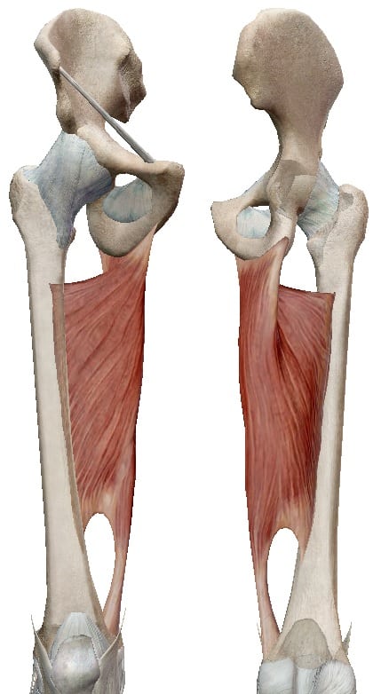
The ischiotibial part goes from its origin to its insertion in a practically vertical path (1, 2).
Actions (1, 2):
- Adduction of the hip joint.
- Flexion of the hip joint (adductor portion).
- Extension of the hip joint (ischiotibial portion).
- Internal rotation of the hip joint.
Gracilis
The gracilis muscle is an elongated leg muscle, the most superficial and internal of the internal compartment of the thigh, and the weakest of the adductor muscles of the hip joint (1, 2).
It is a synergistic muscle in both its actions on the hip joint and the knee joint (2).
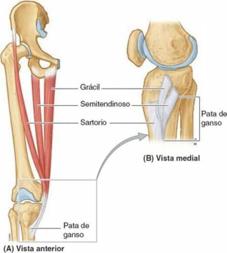
Its insertion on the tibia occurs through a common tendon with the sartorius and semitendinosus muscles (pes anserinus) (1, 2).
- Origin (1, 2)
On the pubis, on the inferior pubic ramus, and on the ischial ramus. - Insertion (1, 2)
On the internal surface of the proximal part of the tibial shaft, between the insertions of the sartorius and semitendinosus muscles (pes anserinus).

Therefore, the gracilis muscle is the only biarticular muscle of the internal compartment of the thigh. From its point of origin, the gracilis muscle follows a practically vertical path to its point of insertion (1, 2).
Actions (1, 2):
- Adduction of the hip joint.
- Flexion of the knee joint.
- Internal rotation of the knee joint.
Posterior compartment of the thigh
The leg muscles found in the posterior compartment form the hamstring muscle group, which are (1, 2):
- Biceps femoris
- Semitendinosus
- Semimembranosus.
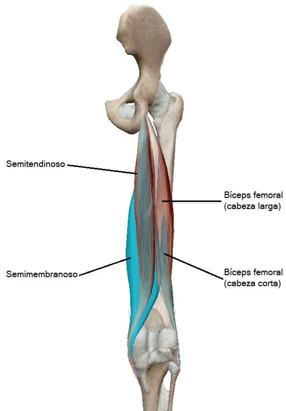
However, since the insertion points of one of these 3 muscles are not on the tibia, it would be more correct to refer to this muscle group as “ischiosural.” This is because the insertion of the biceps femoris muscle occurs on the head of the fibula and not on the tibia (1, 9, 10).
Regarding their main functions, they are extensors of the hip joint and flexors of the knee joint (1, 2), and together they maintain the relaxed upright bipedal position (2).
On the other hand, the hamstring muscles cannot perform their main actions at maximum intensity simultaneously (in full knee flexion, they cannot perform full hip extension and vice versa).
Additionally, the maximum activation of this muscle group occurs in eccentric contractions, that is, decelerating actions contrary to those they perform (2).
Semimembranosus
The semimembranosus muscle is located deeper than the semitendinosus muscle. It is a wide leg muscle whose tendon divides into 3 parts that insert at different points (1, 2)
- Origin (1, 2).
Supero-external impression of the ischial tuberosity. - Insertion (1, 2, 3).
- Groove and contiguous bone of the postero-internal surface of the medial tibial condyle.
- Forms the oblique popliteal ligament (towards the lateral femoral condyle)
- In the popliteal fascia surrounding the knee joint.

Actions (1, 2):
- Flexion of the knee joint.
- Extension of the hip joint.
- Internal rotation of the hip joint (along with the semitendinosus muscle).
- Internal rotation of the knee joint (along with the semitendinosus muscle, when the knee joint is in flexion).
Semitendinosus
The semitendinosus muscle is a fusiform leg muscle, located more internally than the biceps femoris muscle, and ends towards the middle of the thigh, reaching its point of insertion on the tibia through a long tendon. The tendon of the semitendinosus muscle runs the second half of the thigh inferiorly over the semimembranosus muscle (1, 2).
Thus, the name of the semitendinosus muscle is due to the fact that the lower half of its expansion is tendinous (2).
- Origin (1, 2).
Infero-internal part of the upper part of the ischial tuberosity (along with the biceps femoris muscle). - Insertion (1, 2, 3).
Internal surface of the proximal end of the tibia, posterior to the tendons of the gracilis and sartorius muscles, in the pes anserinus.

Actions (1, 2):
- Flexion of the knee joint.
- Extension of the hip joint.
- Internal rotation of the hip joint (along with the semimembranosus muscle).
- Internal rotation of the knee joint (along with the semimembranosus muscle, when the knee joint is in flexion).
Biceps femoris
The biceps femoris muscle is the leg muscle of the posterior compartment of the thigh that inserts into the fibula. It is located on the lateral side of the posterior thigh (1, 2), although one of its heads is oblique (1).
- Origin (1, 2, 3)
- Long head
Inferomedial part of the upper area of the ischial tuberosity. - Short head
Lateral lip of the linea aspera of the femur and lateral supracondylar line of the femur.
- Long head
- Insertion (1, 2)
- Through a common tendon, the biceps femoris muscle inserts into the external surface of the fibular head.
- On the other hand, expansions of this tendon join the lateral collateral ligament and other ligaments of the external side of the knee joint.
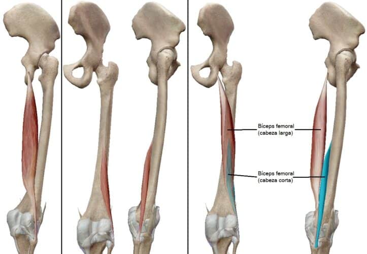
The long head of the biceps femoris muscle is oriented obliquely from its point of origin to its union with the short head in the common tendon (1). Additionally, just inferior to the gluteal region, the long head of the biceps femoris muscle crosses and protects the sciatic nerve (2).
Actions (1, 2):
- Flexion of the knee joint.
- External rotation of the knee joint (when the knee joint is in flexion).
- Extension of the hip joint (long head).
Leg
The leg muscles are located in 3 compartments, one posterior, another anterior, and another lateral (1, 2).
Due to their insertions, the leg muscles can act on the knee joint, on the ankle joint, on the foot, on the toes, or on more than one of these joints and articular complexes (1).
Posterior compartment of the leg
[article ids=”52986″] The posterior compartment of the leg is the largest of the 3 (2) and the leg muscles located in the posterior compartment mainly perform plantar flexion and inversion of the foot and flexion of the toes (1, 2).
These muscles are found in 2 different planes, one deep and one superficial (1, 2), with the superficial plane being the largest in volume (2).
Deep group of the posterior compartment
In this group, there are 4 leg muscles, of which 1 acts on the knee joint, 3 on the ankle joint complex, and 2 of the latter also on the foot (1, 2, 3).
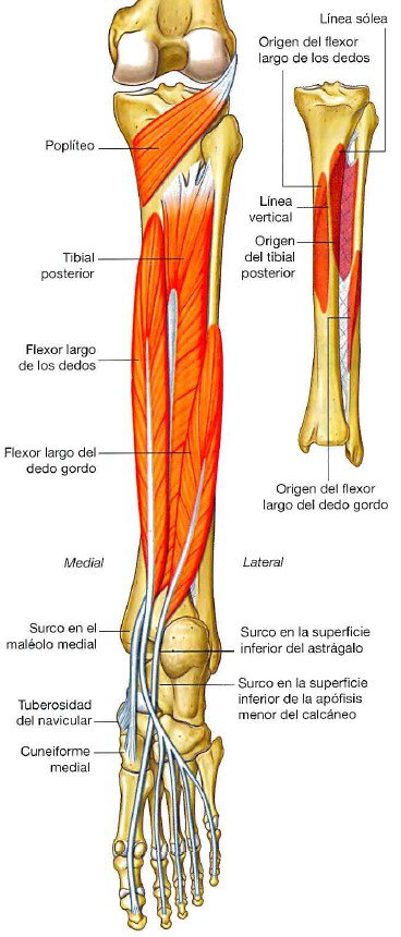
Posterior tibialis
The tibialis muscle is located between the long flexor muscles of the big toe and the long flexor of the toes and is the deepest of the posterior compartment (1, 2). It is one of the most important leg muscles in maintaining the internal arch of the foot during walking (2).
- Origin (1, 3).
Posterior surface of the interosseous membrane of the leg and adjacent surfaces of the tibia (inferior to the soleal line) and the fibula. - Insertion (1, 2, 3, 6)
Plantar surfaces of the bones:
- Tuberosity of the navicular.
- Cuboid.
- Cuneiforms.
- Bases of metatarsals II, III, and IV.
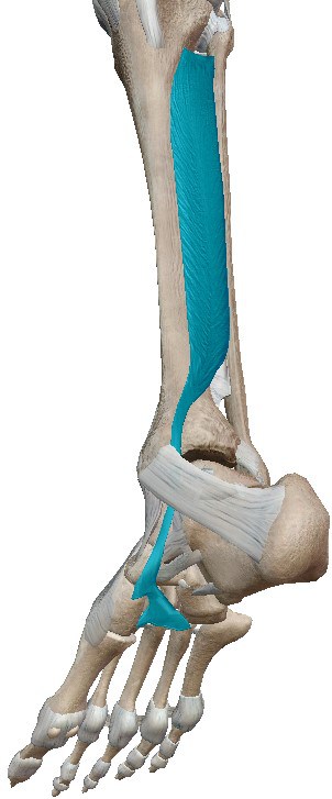
The tendon of the posterior tibialis muscle, in the posterior groove of the medial malleolus, is located internally to the tendon of the long flexor muscle of the toes, where it curves forward to enter the internal side of the foot and surrounds the internal margin of the foot before reaching its insertion points.
Actions (1, 2, 3)
- Inversion of the foot.
- Extension of the ankle joint (plantar flexion).
- Support of the internal arch of the foot during walking.
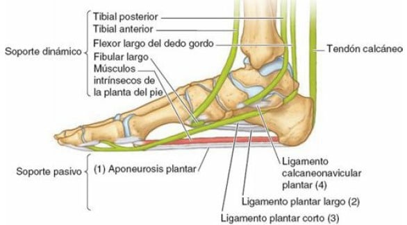
Popliteus
It is a small muscle (1, 3), in fact, the smallest of the posterior compartment of the leg, and is located superior to the other 3 muscles of the posterior compartment (1).
It is a flat, triangular-shaped leg muscle that constitutes part of the floor of the popliteal fossa (1, 2).
- Origin (1, 2)
On the posterior surface of the proximal end of the tibia, between the triangular region and the soleal line. - Insertion (1, 2)
On the infero-external surface of the lateral femoral condyle and on the lateral meniscus.
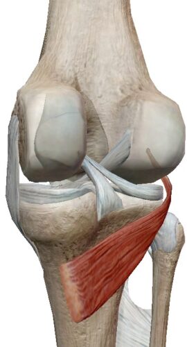
Thus, it is seen how the popliteus muscle follows an ascending and lateral path from its point of origin to its point of insertion, crossing the articular capsule (1, 3, 6), passing between the lateral meniscus and the fibrous membrane (1).
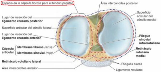
Actions (1, 2, 3):
- Pulling back the lateral meniscus during knee joint flexion.
- Unlocking the knee joint. In standing, it externally rotates the femur on the tibia (internal knee rotation) to initiate flexion.
- Prevents anteriorization of the femur on the tibia when the knee joint is in flexion (assists the posterior cruciate ligament).
- Internal rotation of the knee joint in flexion and without ground contact (assists the semimembranosus and semitendinosus muscles).
Long flexor of the toes
The long flexor muscle of the toes has its origin and much of its path in the leg, and its point of insertion in the toes II, III, IV, and V of the foot. Therefore, it is an extrinsic muscle of the foot (1, 2, 3).
- Origin (1, 2)
Internal surface of the posterior surface of the tibia, inferior to the soleal line. - Insertion (1, 2, 3)
Plantar surface of the 3rd phalanx of toes II, III, IV, and V.
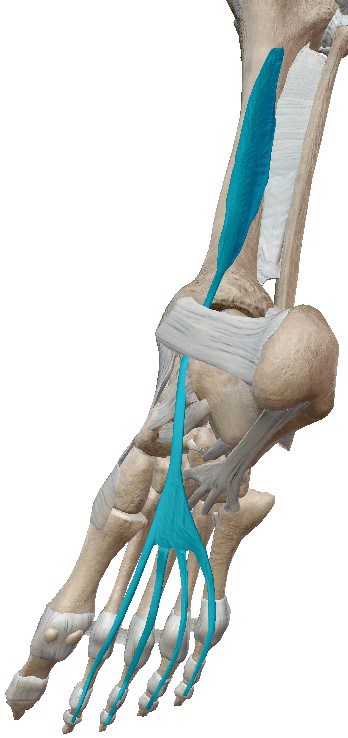
From its point of origin, the long flexor muscle of the toes descends. Near the ankle joint, its tendon follows posterior to the tendon of the posterior tibialis muscle, continues behind the medial malleolus, and on the sole of the foot inferior to the tendon of the long flexor muscle of the big toe, until it reaches its point of insertion (1).
Actions (1, 2, 3):
- Flexion of toes II, III, IV, and V.
- Plantar flexion of the ankle joint complex.
- Support of the longitudinal arches of the sole of the foot.
- Participates in the gripping and propulsion phases of walking.
Long flexor of the big toe
It is an extrinsic leg muscle of the foot (1, 2, 3), meaning it acts on the foot, but at least one insertion point is located in a different area than the foot.
It is a powerful flexor muscle of the big toe (of all its joints) and very important in the final phase of propulsion during walking (2).
- Origin (1, 2, 3)
Posterior surface of the fibula (lower two-thirds) and the interosseous membrane. - Insertion (1, 2, 3)
2nd phalanx of the big toe.
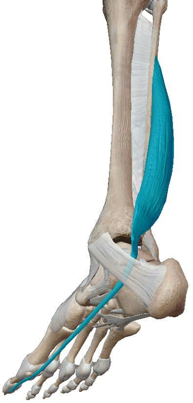
From its point of origin, it descends medially, passing posterior to the distal end of the tibia and through a groove of the talus, curving at 2 points below the talus, and reaching its point of insertion (1, 2).
Actions (1, 2):
- Flexion of all joints of the big toe.
- Plantar flexion of the ankle joint complex (assists).
- Very active in the toe-off phase of the big toe during walking when walking barefoot (if wearing shoes, the final propulsion is part of the plantar flexion performed by the forefoot).
- Support of the internal arch of the foot.
Superficial group of the posterior compartment
They are large muscles (larger than those of the deep group), and powerful, acting in propulsion during walking, running (1, 2), jumping, or walking on the forefoot (tiptoes) (2).
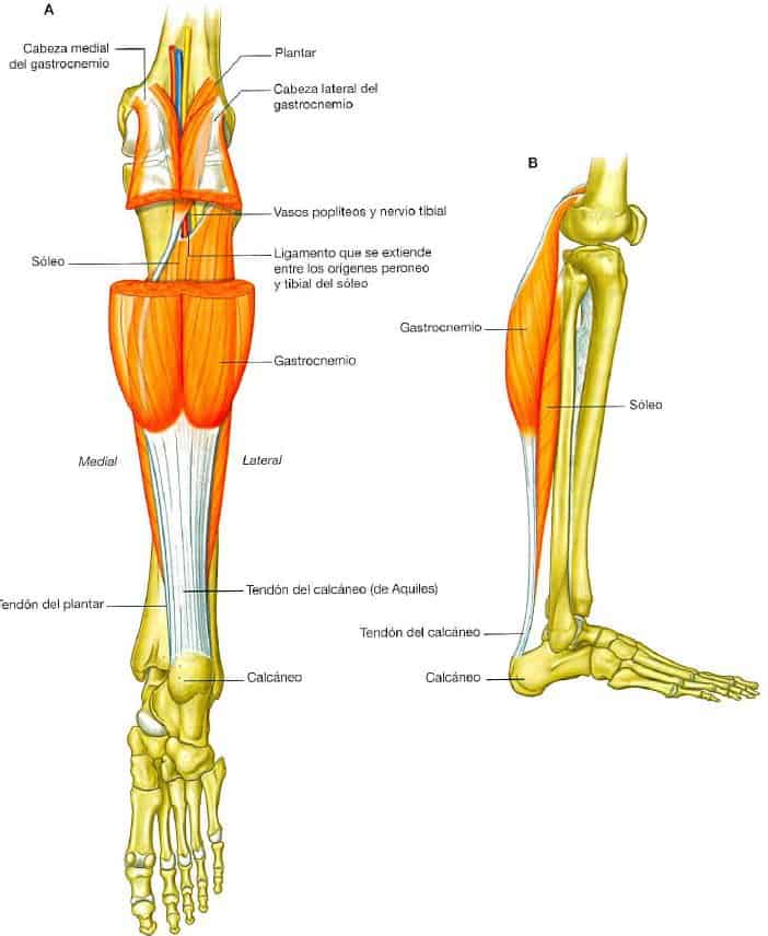
The soleus and gastrocnemius muscles unite in a common tendon (calcaneal tendon/Achilles tendon), forming the 3 heads of the triceps surae (1, 2), a muscle group that can produce up to 93% of the force required in plantar flexion of the ankle joint complex (2).
Triceps surae
The triceps surae is the muscle group formed by 2 leg muscles, the gastrocnemius (medial and lateral) and the soleus muscle (2, 3), which unite in the Achilles tendon, which inserts into the calcaneus (1, 2, 3, 11). The fibers of the triceps surae have a multiplanar arrangement, giving it great power (2, 3).
The common tendon of the triceps surae is a thick and strong tendon (in fact, it is the thickest, most powerful, and resistant in the body) that can reach 15 cm in length (2, 11).
Gastrocnemius
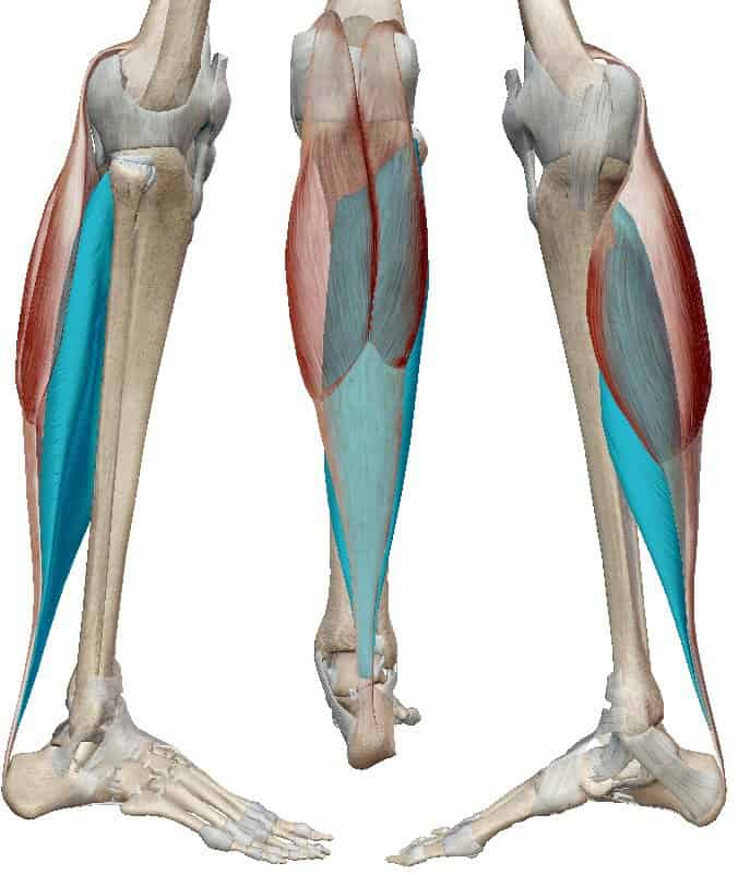
Among the leg muscles, the gastrocnemius is one of the largest and is the most superficial of the posterior compartment (1). It has 2 heads with different points of origin that insert into a common tendon (1, 2, 3). Both heads form the medial and lateral borders of the popliteal fossa and unite to form a single muscle belly (1, 2).
It constitutes the most visible upper part of the calf. The medial head is larger both transversely and longitudinally (2).
Although it is a very powerful biarticular muscle, it cannot act on the knee joint and the ankle joint complex simultaneously, being more effective when the knee joint is in full extension (2).
- Origin (1, 2)
- Medial head
Elongated roughness on the postero-superior surface of the medial femoral condyle and posterior to the adductor tubercle. - Lateral head
Superior and posterolateral surface of the lateral femoral condyle.
- Medial head
- Insertion (1, 2, 3, 11)
On the calcaneal tuberosity through a thick and strong common tendon with the soleus muscle, the calcaneal tendon/Achilles tendon.
Actions (1, 2):
- Extension of the ankle joint complex (plantar flexion) when the knee joint is in extension.
- Flexion of the knee joint.
- Heel elevation during walking.
Soleus
The soleus muscle is a large, flat leg muscle located in a deeper plane than the gastrocnemius muscle (1, 2).
It acts together with the gastrocnemius muscle on this joint complex when the knee joint is in extension and alone when it is in flexion (2).
- Origin (1, 2)
- Soleal line and adjacent medial border of the tibia.
- Posterior surface of the proximal end of the fibula and upper portion of the fibular shaft.
- Soleal arch, between the tibial and fibular insertions.
- Insertion (1, 3, 11)
On the calcaneal tuberosity through a thick and strong common tendon with the gastrocnemius muscle, the calcaneal tendon/Achilles tendon.
Actions (1, 2):
- Extension of the ankle joint complex (plantar flexion).
- Stabilization of the leg in standing.
Plantaris
The plantaris muscle is a small, short (1, 2) fusiform leg muscle located deep to the lateral head of the gastrocnemius muscle, between it and the soleus muscle (1, 2, 12).
The plantaris muscle does not always develop, with 5 to 10% of the population not having it. On the other hand, due to the high density of neuromuscular spindles it has, it has been considered to act as a proprioceptive organ (2).
- Origin (1, 2)
- Lower area of the lateral supracondylar ridge of the femur.
- Oblique popliteal ligament.
- Insertion (1, 2, 11)
Posterior surface of the calcaneus, possibly isolated or attached to the calcaneal tendon/Achilles tendon, fusing with its medial border, almost at the insertion.

External compartment of the leg
In the lateral compartment, it is the smallest of the leg muscle compartments (2) and contains 2 leg muscles that evert the ankle joint (1, 2).
It is located between the external surface of the tibia, the anterior and posterior intermuscular septa (2).
Peroneus brevis
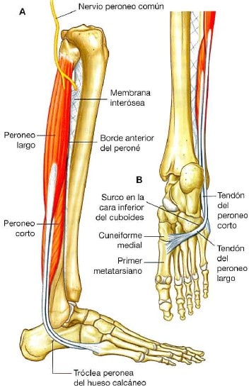
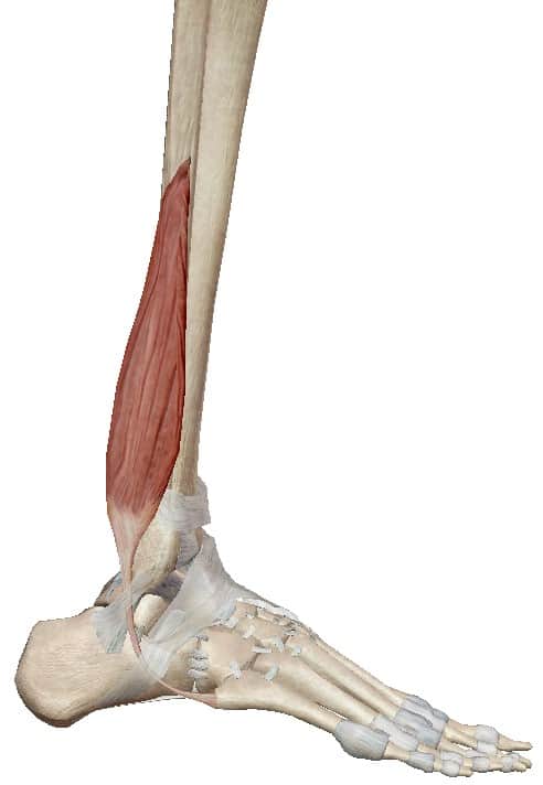
The peroneus brevis muscle is located deeper than the peroneus longus muscle and, like it, passes behind the lateral malleolus, but, on the contrary, inserts on the external side of the foot (1, 2).
- Origin (1, 2)
Distal two-thirds of the external surface of the fibula. - Insertion (1, 2, 3)
External tubercle of the base of the 5th metatarsal.
Actions (1, 2):
- Eversion of the foot (assists).
- Extension of the ankle joint complex (plantar flexion).
Peroneus longus
It is an extrinsic leg muscle of the foot, as it originates in the lateral compartment of the leg, but its insertion occurs on a tarsal bone and a metatarsal bone (1, 2, 3), crossing the Lisfranc joint (3, 13).
- Origin (1, 2)
- Supero-external surface of the fibula.
- Anterior surface of the fibular head and adjacent area of the lateral tibial condyle.
- Insertion (1, 2, 3)
- Infero-external surfaces of the distal end of the medial cuneiform.
- Base of the 1st metatarsal.
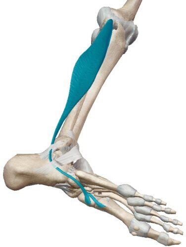
From its point of origin, it passes posterior to the lateral malleolus through a bony groove and is oriented anteriorly to the external area of the foot. Through a groove on the inferior surface of the cuboid, it crosses the sole of the foot internally to its insertion points (1, 2).
Actions (1, 2):
- Eversion of the foot.
- Extension of the ankle joint complex (plantar flexion).
- Together with the anterior tibialis and posterior tibialis muscles, it serves as a “stirrup,” supporting the plantar arches (the peroneus longus especially the external and anterior arches).
Anterior compartment of the leg
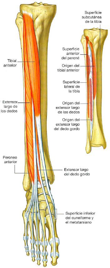
The anterior compartment is located between the external surface of the tibia, the internal surface of the fibula, and anterior to the interosseous membrane (2).
In the anterior compartment, there are 4 leg muscles that are mainly flexors and inverters of the ankle joint complex (dorsiflexion) and extensors of the toes (1, 2).
Long extensor of the toes
The long extensor muscle of the toes is the most posterior and external leg muscle of the 4 muscles of the anterior compartment (1, 2).
- Origin (1, 2)
- Lateral tibial condyle
- Upper half of the internal surface of the fibula.
- Interosseous membrane.
- Deep fascia.
- Insertion (1, 2, 3)
Dorsal surfaces of the 2nd and 3rd phalanges of toes II, III, IV, and V.
<img class=”wp-image-80382 size-full” src=”https://mundoentren
