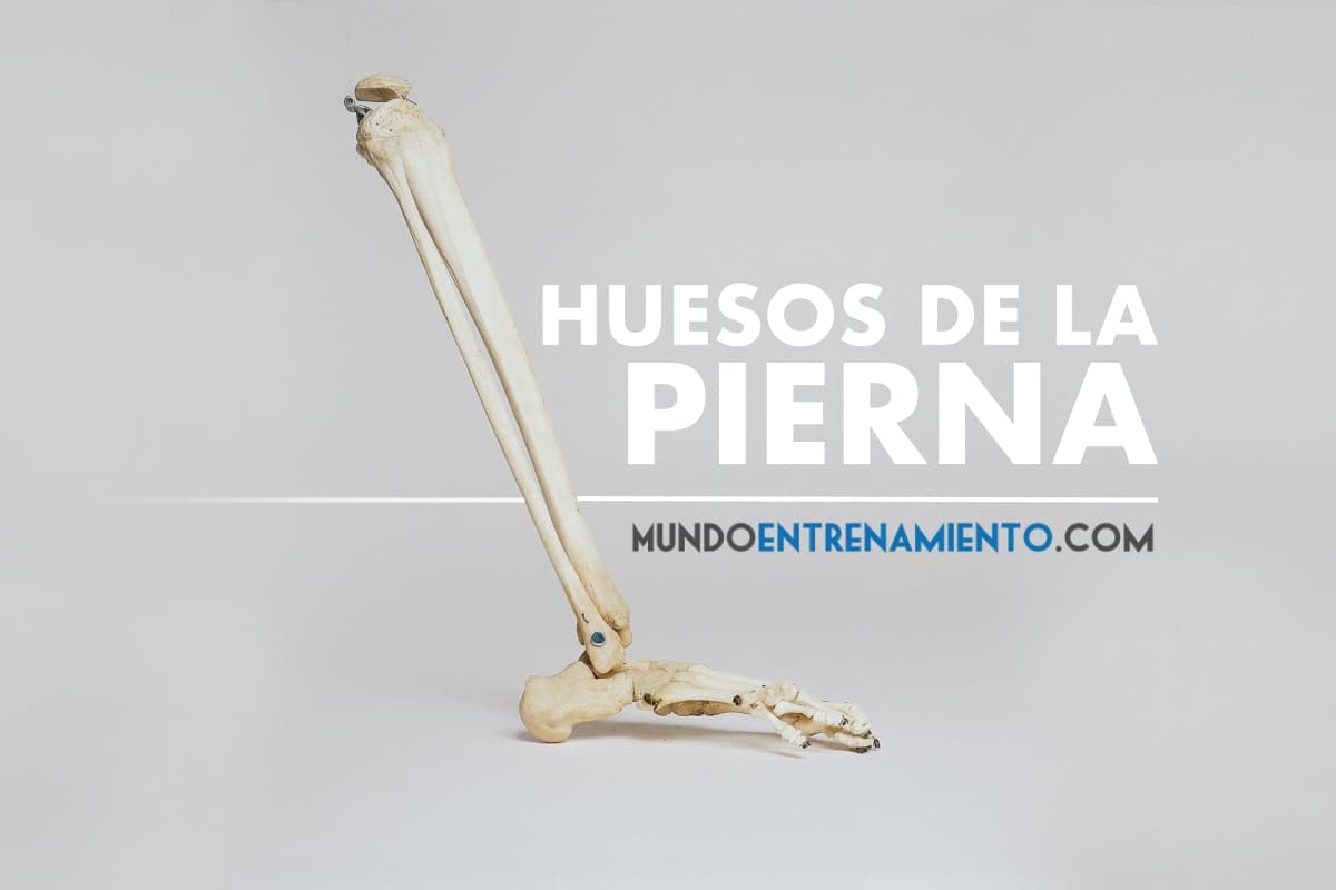
The bones of the leg are a total of 31, 2 that form the hip and thigh joint, 3 that form the leg, and 26 that form the foot.
On the other hand, although colloquially referred to as “leg bones,” anatomically it is called the lower limb, which consists of the hip (joint), thigh, leg, and foot (1, 2, 3, 4, 5).
This article aims to detail in a practical way all the bones of the leg.
What are the bones of the leg? We analyze the different parts of the leg
The lower limb is the extremity whose main function is to ensure bipedalism, for this reason, the bones of the leg, as well as the joints, are larger and present greater stability than the upper limb (5).
Thus, the bones of the leg or parts of the leg (referring to the lower limb) are distributed in different regions:
Pelvis
From the pelvic region, the coxal bone is the point of union, along with the femur, between the lower limb and the trunk (throughout the pelvic region) (1, 2, 5).
The coxal bone is one of the bones of the leg, formed by 3 embryonic bones (ischium, pubis, and ilium) (1, 4), which has a curved relief and whose contours are irregular (1, 2, 5).
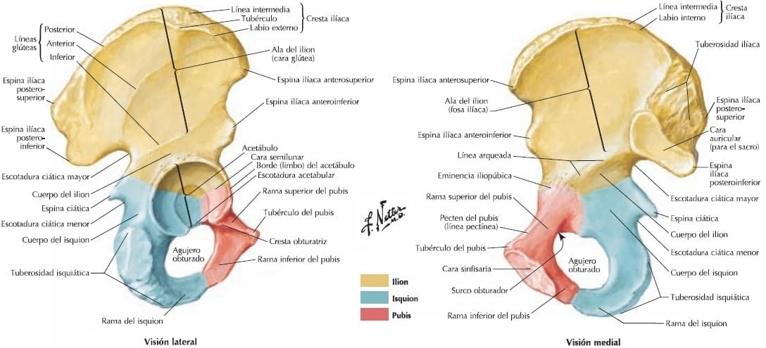
This bone consists of 3 parts: the superior and inferior serve as muscle insertions and to form the pelvis (superior part through the sacroiliac joint and the inferior part through the pubis) (1, 5).
The superior part has a flatter surface called the iliac wing, which on its external face presents the gluteal surface and on its internal face the iliac fossa. (1, 5).
At the cranial (superior) level is the iliac crest, which follows a path from anterior to posterior in an irregular fan shape (1, 2, 5).
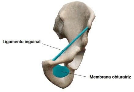
The inferior part presents the obturator foramen, which is almost entirely closed by the obturator membrane (1, 2, 5).
From the anterior superior iliac spine to the pubic tubercle runs the inguinal ligament or crural (1, 2, 6).
Among the bones of the leg, in the middle part, on its external face, it articulates with the femur thanks to the acetabulum, thus forming the hip joint (acetabular cavity-femoral head) (1, 2, 4, 5).
The acetabulum is an incomplete circular cavity at the lower part (acetabular notch) (1, 4).
The depression that presents from the acetabular notch upwards on the surface of the acetabulum is called the acetabular fossa (2, 4), around which is the lunate surface, which is the articular surface of the coxal in the hip joint (1, 2, 4).
Thigh
The thigh is the strongest part of the lower limb (5), formed at the bone level by the femur bone (1, 2, 5).
Of the bones of the leg (lower limb) and the entire body, the femur is the largest (4, 5) and heaviest bone, reaching a quarter of a person’s height (4).
In the lower image, we see the femur, one of the bones of the leg:
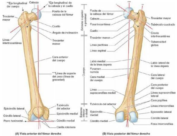
The femur consists of a body (diaphysis) and two ends (epiphyses), one proximal and one distal (4, 5).
In the proximal epiphysis are the femoral head, the femoral neck, the greater trochanter, and the lesser trochanter (the trochanters are bony protrusions, in which several muscle insertions occur (1, 2, 4, 5).
The femoral head is semi-spherical in shape (1, 2, 4, 5) (3/4 of a sphere (4)) with a depression at the medial level, called the fovea of the femoral head (1, 2, 4), from which originates the ligament of the femoral head (1, 2). The femoral head is covered by articular cartilage except for the fovea of the femoral head (1, 2, 4).
The femoral neck is trapezoidal in shape (4). The narrow part (proximal) supports the femoral head and the wider part continues with the diaphysis (1, 2, 4).
At the proximal level, the femur is “L” shaped, so that the axis of the proximal epiphysis is oblique with respect to the body (1, 2, 4).
The angle in adults is 115-140º, usually smaller in women than in men (due to greater width of the acetabular cavity and obliquity of the femoral head) (4) causing the femur to have an oblique disposition in an inferomedial direction (1, 2, 4, 5).
This makes it possible for the knees to be located under the trunk, making bipedal walking possible, by projecting the center of gravity towards the vertical axes of the legs (4).
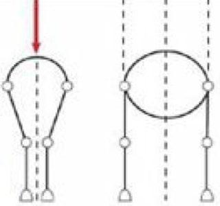
Above the junction of the proximal epiphysis and the diaphysis, 2 protrusions arise: the greater trochanter and the lesser trochanter (1, 2, 4, 5).
The greater trochanter, is one of the bones of the leg and is located laterally and goes superiorly and posteriorly at the epiphysis-diaphysis junction, protruding medially from a posterior and/or superior perspective over the trochanteric fossa.
The lesser trochanter, another of the bones of the leg, is conical in shape with a rounded tip and goes from the posteromedial part of the epiphysis-diaphysis junction towards medial (1, 2, 4).
The mentioned junction point between the epiphysis and the diaphysis can be seen from an anterior view by the intertrochanteric line, a rough crest that goes from the greater trochanter to surround the lesser trochanter and, from a posterior view, by the intertrochanteric crest, which is more prominent (1, 2, 4).
On the intertrochanteric crest, there is a prominence called the quadrate tubercle (1, 4).
The diaphysis of the femur is mostly smooth and rounded, with its anterior part somewhat convex.
On its posterior part, it has a wide, rough, and longitudinal line called the linea aspera, which has 2 borders, the lateral lip, which superiorly joins the gluteal tuberosity, and the medial lip that continues medially and superiorly (1, 2, 4) as the spiral line (4).
Between the medial lip and the spiral line, below the lesser trochanter, is the pectineal line (1, 2, 4), derived from the linea aspera. Inferiorly, the linea aspera divides into the medial supracondylar line and the lateral supracondylar line, which reach the medial condyle and lateral condyle respectively (1, 4).
The femoral condyles are joined anteriorly and separated inferiorly and posteriorly by the intercondylar fossa. On the anterior part, they form the patellar surface (1, 2, 4, 5) or trochlea (5).
Both condyles are similar, but the medial condyle is longer and narrower (1, 2, 4, 5).
The 2 present an eminence, the lateral condyle on its lateral face and the medial condyle on its medial face, called epicondyles (lateral and medial respectively), and above the medial epicondyle is the adductor tubercle (1, 2, 4).
Knee
The knee is the joint of the femur and tibia, but there is a third bone involved in this joint, which does not belong to either the thigh or the leg:
the patella, is one of the bones of the leg (1, 2, 3, 4, 5, 7) or patella, by its English name (7), and understanding the knee as an anatomical region, it is the region to which this bone belongs (1, 2, 3, 4, 5, 7).
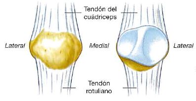
The patella is considered a sesamoid bone (3, 4, 7, 8), a type of bone that develops in some tendons that act on a joint (3, 4, 9).
It is the largest sesamoid bone in the body (3, 4), whose function is to protect the anterior face of the knee joint and also the quadriceps muscle, as well as to improve the efficiency of this muscle (8).
It is located within the quadriceps tendon, in front of the knee (1, 2, 3), between the quadriceps tendon (within which it is located) and the patellar ligament (1, 2, 3, 4) or patellar tendon, as it is the continuation of the quadriceps tendon (3, 4, 10).
It is triangular in shape, with the apex at its lower part (2, 3, 7).
The base is superior, wide, and thick (2, 3), and the posterior face, the articular face (1, 2, 3) presents 2 facets, one lateral and another medial, separated by a central crest (1, 2, 3).
The lateral facet is larger (1, 3), due to the larger surface of the trochlea corresponding to the lateral condyle (1, 2, 3).
Leg
The leg is the region of the lower limb that is between the thigh and the foot. The 2 bones of the leg are the tibia and the fibula (1, 2, 3, 4, 5).
Tibia
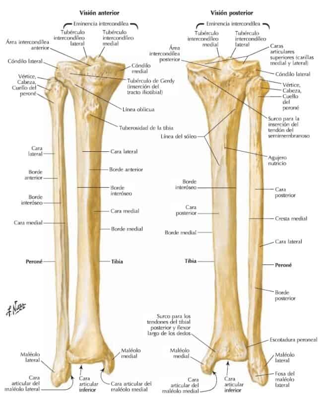
Of the 2 bones of the leg, the tibia is the largest (3) and thickest (5) bone of the leg and is responsible for transmitting the weight of the femur to the foot (4, 5).
At the proximal level, it presents the tibial plateau, an articular face formed by the medial condyle, the lateral condyle, and an intercondylar region that separates them.
The intercondylar region is formed, in its central part, by the intercondylar eminence, formed in turn by the medial intercondylar tubercles and lateral intercondylar tubercles, and in its anterior and posterior parts by the anterior intercondylar areas and posterior intercondylar areas (1, 2, 3, 4).
The tibial condyles are concave at the superior level, accentuating this concavity towards medial, and flatter by their external edge (3).
The intercondylar region presents 6 facets that serve as insertion for ligaments and menisci, arranged from anterior to posterior.
The lateral condyle presents at the antero-external level, and inferior to the articular face, the Gerdy’s tubercle (1, 4), and at the postero-external and inferior level, the fibular articular face (3, 4, 5)
The diaphysis of the tibia is arranged vertically (4) and presents 3 faces (lateral, medial, and posterior) and 3 borders (anterior, medial, and interosseous) (1, 2, 3, 4).
At the upper part of the anterior border, below the junction of both tibial condyles, is the tibial tuberosity, a wide triangular-shaped prominence with the apex oriented inferiorly, which serves as the insertion point for the patellar ligament (1, 2, 3, 4).
At the proximal end (internally with respect to the tibial tuberosity) the medial face presents a more elevated and somewhat rough area (3). The medial face is smooth (1, 2, 3, 4) and like the anterior border (4), subcutaneous (3).
The posterior face is wider at its upper part and is traversed obliquely from supero-external to infero-internal to the medial border by the soleal line (1, 2, 3, 4).
The distal end of the tibia is smaller than the proximal, and extends on its medial part to form the medial malleolus below the body (1, 2, 3, 4).
On the posterior part is the malleolar groove, which is oriented inferiorly and medially to the medial malleolus (3) and on the lateral surface, at the distal end of the interosseous border, is the fibular notch or peroneal notch, an articular face for the fibula.
Fibula
The fibula is the smaller bone of the 2 bones of the leg (region of the lower limb) (1, 2, 3, 4) and its function is not to support body weight (3, 4), but mainly to serve as a muscle insertion both distally and proximally (4).
At the proximal end of the fibula (another of the bones of the leg) is the head of the fibula (1, 2, 3, 4), on whose supero-internal face is a circular facet (3) with which it articulates with the tibia and a pointed apex (styloid process), located postero-laterally to the articular facet (1, 2, 3, 4).
Like the tibia (another of the bones of the leg), the diaphysis of the fibula presents 3 borders (anterior, interosseous, and posterior) and 3 faces (medial, lateral, and posterior) (1, 2, 3, 4). The posterior face is vertically divided into 2 parts by the medial crest (1, 2, 3).
The distal end grows postero-inferiorly, forming the lateral malleolus (1, 2, 3, 4, 5) which is more posterior and inferior than the medial malleolus of the tibia (1, 2, 4, 5).
At the distal end of the fibula is an articular facet on its medial part (ankle joint complex) (1, 2, 3, 4), above which is a triangular face (3) without cartilage (5) for the tibiofibular syndesmosis (joint of the tibia and fibula whose surfaces are joined by a ligament or membrane) (3, 5, 11).
Postero-inferiorly is the malleolar fossa (1, 2, 3). On the posterior surface of the lateral malleolus is the malleolar groove (2, 3).
Foot
The foot is the most distal region of the lower limb and where 26 of the 31 bones of the leg (lower limb) are found (1, 2, 3, 4, 5).
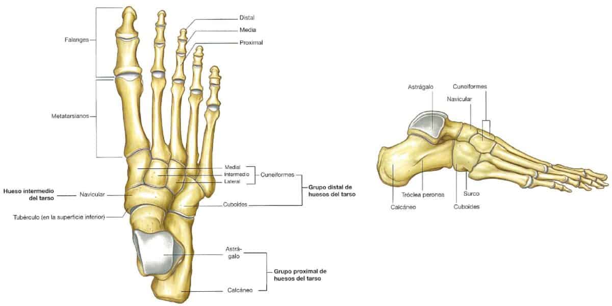
The foot is divided into 3 zones (1, 2, 3, 4, 5):
- Tarsus (7 bones).
- Anterior tarsus (5 bones).
- Posterior tarsus (2 bones).
- Metatarsus (5 bones).
- Phalanges (14 bones).
In the proximal group of the tarsus (posterior tarsus), which corresponds to the hindfoot (4), are the talus and the calcaneus.
The talus is a small bone, supported on the calcaneus and articulates with the tibia and fibula forming the ankle, so it is responsible for receiving the body’s weight (1, 2, 3, 4, 5).
The talus has a body, neck, and head (1, 2, 3, 4) which is anterior to the tibia (1, 2, 3, 5) and articulates with the navicular bone (or tarsal scaphoid) (1, 2, 3).
No muscles insert on the talus (1, 4) and it has articular cartilage on almost its entire surface (2, 3, 4) to articulate superiorly or medially with the tibia, laterally with the fibula, anteriorly with the navicular, and inferiorly with the calcaneus (1, 2, 3, 4).
The calcaneus is posterior to the ankle forming the structure of the heel (1, 2, 3, 4, 5). It is the foot bone that bears the most weight (4). Its posterior part, rounded, is divided into superior, middle, and inferior, with the inferior bearing the weight (3).
The navicular is the intermediate bone of the tarsus (3) and along with the bones of the leg, of the distal group (anterior tarsus) correspond to the midfoot (3, 4).
Its medial face presents the navicular tuberosity (1, 2, 3, 4), located on the medial edge of the foot, which forms the plantar arch (4, 5).
In the distal group of the tarsus (anterior tarsus) are the cuboid bone, and the 3 cuneiform bones or wedges (medial, intermediate, and lateral or 1st, 2nd, and 3rd) (1, 2, 3, 4, 5).
The cuboid is the lateral bone of the anterior tarsus and articulates posteriorly with the calcaneus and anteriorly with the 2 most lateral metatarsals (1, 2, 3, 4).
Infero-laterally it presents the cuboid tuberosity and anterior to this, on the plantar surface, the groove of the peroneus longus tendon (1, 2, 4).
The 3 cuneiform bones articulate with each other, posteriorly with the navicular, anteriorly with the respective metatarsal, and in the case of the lateral cuneiform, with the cuboid (1, 2, 3, 4).
The metatarsus, along with the phalanges, constitutes the forefoot, the anterior half of the foot (4). The metatarsus is formed by 5 metatarsal bones, numbered I-V from medial to lateral (1, 2, 3, 4, 5).
These bones of the leg are long bones, so they have a base (proximal end), a diaphysis, and a head (distal end).
Each metatarsal base articulates with the cuneiform bones and the cuboid, and those of metatarsals II-V with each other. On the other hand, each metatarsal head articulates with the proximal phalanx of the corresponding toe (1, 2, 3, 4, 5).
The first metatarsal is the shortest and strongest, and the second the longest (3, 4).
The bases of the I and V metatarsal present tuberosities for tendon insertions (4), and the tubercle of the V metatarsal is oriented postero-laterally (1, 2, 3, 4).
The head of the I metatarsal, on its plantar face, articulates with 2 sesamoid bones (medial and lateral) (1, 2, 3, 4, 5), included in the tendon of the short flexor muscle of the 1st toe (1), which slide through 2 grooves present on the head of the I metatarsal (5).
There are a total of 14 phalanges in each foot (1, 2, 3, 4), 3 for each toe from the 2nd to the 5th (proximal, middle, and distal) and 2 for the 1st toe (proximal and distal) (1, 2, 3, 4, 5).
These bones of the leg are small long bones, so they also have a base (proximal end), body (diaphysis), and head (distal end) (1, 2, 3, 4).
The bases of the proximal phalanges articulate with the corresponding metatarsal and the heads with their middle phalanx (1, 2, 3, 4).
However, the heads of the distal phalanges do not articulate, but flatten forming a plantar tuberosity in a crescent shape (1, 2, 3).
Interactive game to learn the bones of the human body
Below we leave you an activity to interact and easily locate the main bones of the human body. Let’s see how many you get right!
Conclusion
The bones of the leg have a varied morphology and as already seen in this article, their main function is to support body weight and maintain a bipedal posture.
For this reason, and as Dufour (2012) (5) highlights, the bones of the leg are more robust than those of the upper limb, despite the fact that the bones of the leg (lower limb) and the bones of the upper limb are homologous.
Bibliographic references
- Netter F.H.. (2011). Atlas of Anatomy. 5th edition. Barcelona: Elsevier (Masson publication).
- Staubesand J. (1991). Sobotta. Atlas of human anatomy. Volume 2. Thorax, abdomen, pelvis, and lower limb. 19th edition. Madrid: Médica Panamericana
- Drake R.L., Vogl W., Mitchell A.W.M. (2005) Gray. Anatomy for students. 1st edition. Madrid: Elsevier Spain.
- Moore K.L., Dailey A.F., Agur A.M.R. (2013). Clinically oriented anatomy. 7th edition. Barcelona: Wolters Kluwer/Lippincott Williams & Wilkins Health.
- Dufour M.. (2012). Anatomy of the lower limb. EMC – Podiatry; 14(4), 1-12 [article E – 27-005-A-10]. https://doi.org/10.1016/s1762-827x(12)61929-4
- Clínica Universidad de Navarra (2020) Clínica Universidad de Navarra: Medical Dictionary. Inguinal Ligament. Pamplona, Spain: Medical Dictionary. Retrieved from: https://www.cun.es/diccionario-medico/terminos/ligamento-inguinal. Consulted on 03/12/2020.
- Real Academia Nacional de Medicina de España. (2012) Dictionary of Medical Terms. Patella. Madrid, Spain: Médica Panamericana. Real Academia Nacional de Medicina de España. Dictionary of Medical Terms. Retrieved from: https://dtme.ranm.es/buscador.aspx?NIVEL_BUS=3&LEMA_BUS=rotula. Consulted on 10/12/2020.
- Sherry E., Wilson S.F. (2002) Oxford Manual of Sports Medicine. 1st edition. Barcelona: Paidotribo.
- Real Academia Nacional de Medicina de España. (2012) Dictionary of Medical Terms. Sesamoid Bone. Madrid, Spain: Médica Panamericana. Real Academia Nacional de Medicina de España. Dictionary of Medical Terms. Retrieved from: https://dtme.ranm.es/buscador.aspx?NIVEL_BUS=3&LEMA_BUS=hueso%20sesamoideo. Consulted on 10/12/2020.
- Real Academia Nacional de Medicina de España. (2012) Dictionary of Medical Terms. Patellar Tendon. Madrid, Spain: Médica Panamericana. Real Academia Nacional de Medicina de España. Dictionary of Medical Terms. Retrieved from:. https://dtme.ranm.es/buscador.aspx?NIVEL_BUS=3&LEMA_BUS=tendon%20rotuliano. Consulted on 10/12/2020.
- Real Academia Nacional de Medicina de España. (2012) Dictionary of Medical Terms. Syndesmosis. Madrid, Spain: Médica Panamericana Real Academia Nacional de Medicina de España. Dictionary of Medical Terms. Retrieved from:. https://dtme.ranm.es/buscador.aspx?NIVEL_BUS=3&LEMA_BUS=sindesmosis. Consulted on 04/01/2021.
